A Rare Condition of Vertebral Squaring Accompanied by Hereditary Spherocytosis
By Ayca Uran San1, Ahmet Tezce2, Hatice Gulsah Karatas2, Betul Ustun2, Ramazan Gunduz2, Mufit Akyuz2Affiliations
doi:ABSTRACT
Vertebral squaring is a radiological term which defines straightening of the ventral section of vertebral body. This condition generally occurs as a result of rheumatological inflammation such as ankylosing spondylitis. There are also several other clinical conditions rarely giving rise to this change, like Paget’s disease, inflammatory arthritis, and Down’s syndrome.
To the best of our knowledge, this is the first case of vertebral squaring accompanied by hereditary spherocytosis (HS) in literature. HS is a relatively rare condition of hemolytic anaemia due to red cell membrane defects.
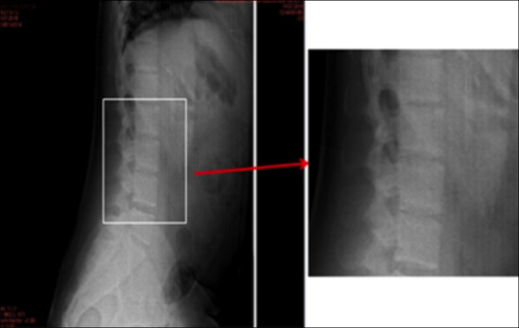
A 28-year male was admitted to our department with low back pain. The patient had been diagnosed with HS. On radiological examination, vertebral squaring was detected by lateral radiograph and MRI of the lumbar spine.
The detection of vertebral squaring is often confusing as the clinical picture may mimic other pathologies. The diagnosis should be confirmed by laboratory and radiological assessments.
Key Words: Anaemia, Spine, Bone marrow.
INTRODUCTION
Hereditary spherocytosis (HS) is a relatively rare condition and is the most common form of hemolytic anaemia caused by defects in red blood cell membrane.1 HS occurs due to a deficiency of ankyrin and spectrin proteins in red blood cell membranes and can be inherited in both autosomal dominant and autosomal recessive patterns.2 Laboratory findings such as anaemia, increased mean corpuscular haemoglobin concentration, microspherocytes, reticulocytosis, and hyperbilirubinemia are characteristic of HS.3 This study presents a case of a patient with HS who was investigated with a preliminary diagnosis of spondyloarthropathy accompanied by the detection of vertebral squaring.
CASE REPORT
A 28-year- male was admitted to our department with low back pain. His medical history revealed that he had inflammatory pain and morning stiffness for 2 months.
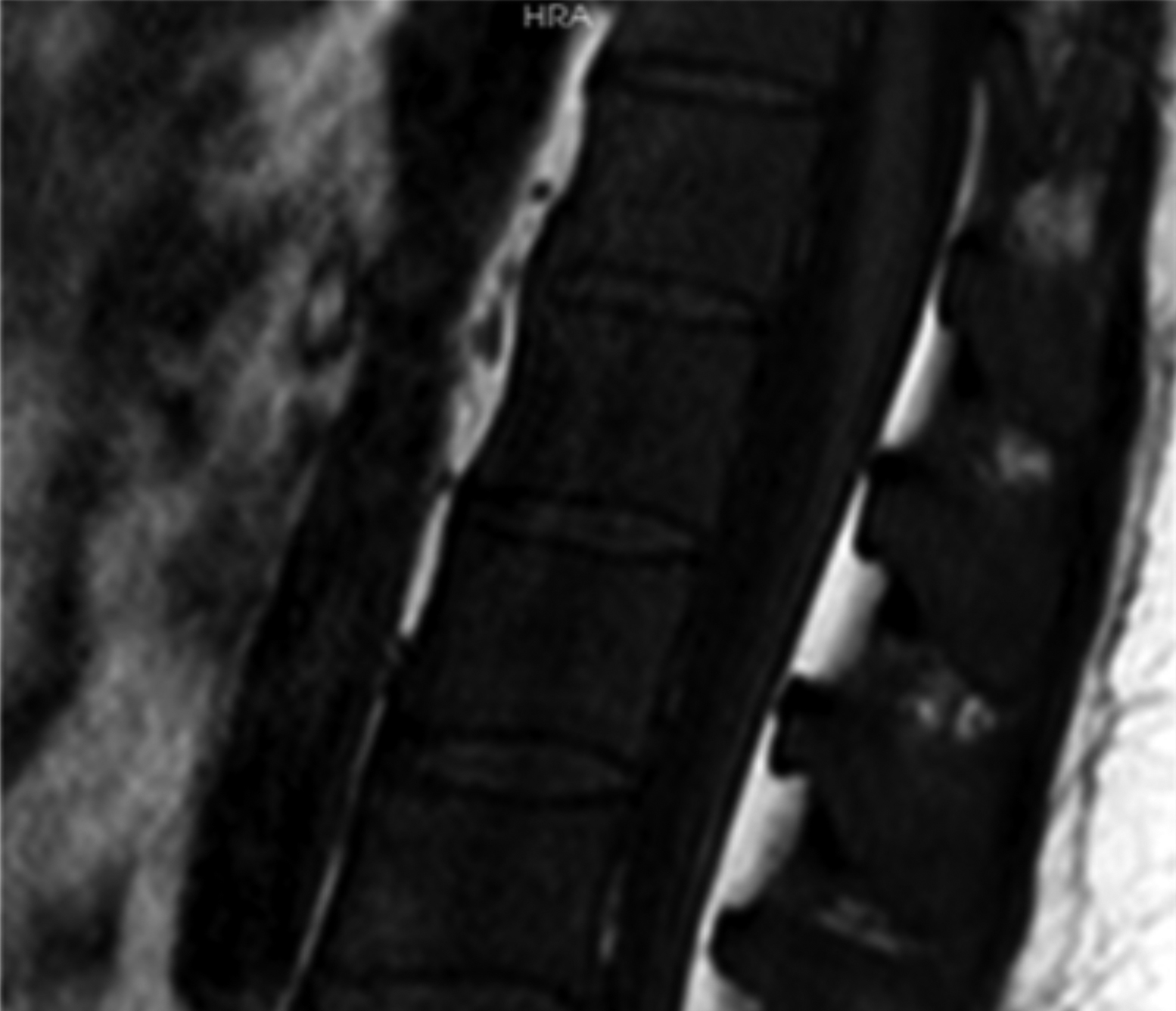
The patient had been diagnosed with HS previously and also had a splenectomy operation 13 years ago. He was receiving depocilin treatment bimonthly. Physical examination revealed that the range of motion (ROM) of all the joints was within normal limits; however, the patient felt pain during the ROM examination of the low back. Tenderness was also detected over the spinous processes of lumbar column by palpation. The straight leg raise (SLR) test was negative. Modified Schober test and chest expansion were in normal ranges. The results of the flexion abduction external rotation (FABER) and flexion adduction internal rotation (FADIR) tests were negative. The bilateral sacroiliac compression tests were evaluated as positive. No sensory-motor deficits were observed. Laboratory findings revealed haemoglobin of 10.1 g/dL, white blood cell count, 17.38 × 10⁹/L, mean corpuscular volume, 109 fL, platelet count, 664.000/mm3, serum C-reactive protein, 10.6 mg/L, erythrocyte sedimentation rate, 10 mm/1st h, and HLA B-27 was negative. The radiograph of the pelvis did not show any irregularity in the bilateral sacroiliac joints. However, there was vertebral squaring on the lateral radiograph of the lumbar spine (Figure 1). Magnetic resonance imaging (MRI) of the lumbar spine was also compatible with vertebral squaring (Figure 2) and revealed an increased signal intensity of the vertebral bodies on the T2-weighted images. The MRI scans of the sacroiliac joints were reported as normal. The patient had consulted the department of chest diseases for his cough and high blood cell count. He was diagnosed with pneumonia and the chest-disease specialist planned antibiotherapy for the patient. Additionally, a physical therapy program including hotpack, transcutaneous electrical nerve stimulation, ROM, stretching, and progressive resistance exercises were also planned for the patient after his recovery from the pulmonary infection. The patient was also administered diclofenac sodium treatment as 50 mg twice a day. After the treatment, the visual analog scale (VAS) pain score decreased from 100 mm to 0 mm according to the assessments at baseline, 3 months, 6 months and 1 year after the treatment.
 Figure 1: The lateral radiograph of the lumbar spine showing vertebral squaring.
 Figure 2: The MRI of the lumbar spine was also compatible with vertebral squaring.
DISCUSSION
Vertebral squaring is a radiological term that defines the straightening of the ventral section of the vertebral body.4 This phenomenon generally occurs as a result of rheumatological inflammation such as ankylosing spondylitis.4 The pathogenesis of vertebral squaring is thought to be related to incomplete formation of new cortex and spongiosa in the vertebrae due to the primary inflammatory reaction in ankylosing spondylitis.5 Additionally, there are several clinical conditions, such as Paget’s disease, inflammatory arthritis, and Down’s syndrome that are rarely accompanied by vertebral squaring.6,7 To the best of our knowledge, this study is the first to report a patient with vertebral squaring accompanied by HS.
Chronic anaemias give rise to functional demand for hematopoiesis which leads to modifications in the bone marrow.6 These alterations result in various side effects on the musculoskeletal system.6
A study has reported the development of high-grade osteoporosis, cortical thinning in the spine, increased height-to-width ratio of the vertebral body (due to medullary expansion), biconcave / wedge-shaped vertebrae and compression fractures related to marrow hyperplasia in patients with thalassemia.6Additionally, another study has detected that a scalloped cortex edge occurs in extremities due to growing extramedullary hemopoietic tissues beneath the periosteum.7
In this case, we hypothesize that vertebral squaring occurred due to the hematopoietic process of HS.
Sclerosis and fibrotic changes lead to infarction in cortical sequences along with marrow and medullary trabeculae; therefore, there is an occurrence of dactylitis which is a clinical condition including fever, and painful hand and feet.6,8,9
This patient was prediagnosed with spondyloarthropathy because of the presence of inflammatory low back pain and detection of vertebral squaring. However, this clinical condition was ruled out based on the laboratory and MRI findings. Finally, we concluded that the patient experienced low back pain due to a lumbar muscle spasm, which occurred as a result of the patient’s complaint of cough.
The objective of this case report was to emphasise that increased hematopoiesis should be considered in the differential diagnosis of vertebral squaring. The detection of vertebral squaring, especially with low back pain, is often confusing because the clinical picture may mimic other pathologies such as spondyloarthropathies. Therefore, the diagnosis should be confirmed by laboratory and radiological assessments such as radiography and MRI.
PATIENT’S CONSENT:
The informed consent is obtained from the patient to publish the data concerning this case.
COMPETING INTEREST:
The authors declared no conflict of interest.
AUTHORS’ CONTRIBUTION:
AUS: Concept, data collection, material, analysis and interpretation, literature review, critical review, and drafting.
AT, BU: Data collection, material, analysis and interpretation, and drafting.
HGK, RG, MA: Concept, design, supervision, and critical review.
All the authors have approved the final version of the manuscript to be published.
REFERENCES
- King MJ, Zanella A. Hereditary red cell membrane disorders and laboratory diagnostic testing. Int J Lab Hematol 2013; 35(3):237-43. doi: 10.1111/ijlh. 12070.
- Koju S, Makaju R. Hereditary Spherocytosis: A case report. J Lumbini Medical College 2018; 6(1):41-3. doi: 10.22502/ jlmc.v6i1.202.
- Sheikh MK, Yusoff NM, Kaur G, Aziz Khan F. Hereditary spherocytosis in a malay patient with chronic haemolysis. Malays J Med Sci 2007; 14(2):54-7. PMID: 22993492; PMCID: PMC3442627.
- Lingg GM, Schorn CS. Square vertebrae and barrel‐shaped vertebrae. In: Baert AL, editor. Encyclopedia of diagnostic ımaging. Berlin (Germany): Springer 2008; p.1733-8. doi.org/10.1007/978-3-540-35280-8_2366.
- Aufdermaur M. Pathogenesis of square bodies in ankylosing spondylitis. Ann Rheum Dis 1989; 48(8): 628-31.8. doi: 10.1136/ard.48.8.628.
- Martinoli C, Bacigalupo L, Forni GL, Balocco M, Garlaschi G, Tagliafico A. Musculoskeletal manifestations of chronic anemias. Semin Musculoskelet Radiol 2011; 15(3):269-80. doi: 10.1055/s-0031-1278426.
- Perisano C, Marzetti E, Spinelli MS, Maria Calla CA, Graci C, Maccauro G. Physiopathology of bone modifications in beta-thalassemia. Anemia 2012; 2012:320737. doi: 10.1155/ 2012/320737.
- Madani G, Papadopoulou AM, Holloway B, Robins A, Davis J, Murray D. The radiological manifestations of sickle cell disease. Clin Radiol 2007; 62(6):528-38. doi: 10.1016/j. crad.2007.01.006.
- Lonergan GJ, Cline DB, Abbondanzo SL. Sickle cell anemia. Radiographics 2001; 21(4):971-94. doi: 10.1148/radio graphics.21.4.g01jl23971.